
When we talk about the nervous system, we almost automatically talk about neurons. They receive signals, generate action potentials, form synapses, and participate in learning, memory, movement, and consciousness. Yet for a long time, another vast cellular world remained largely in the background of that story — neuroglia.
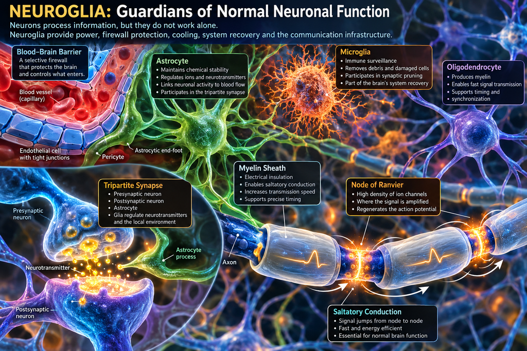
The word glia itself means “glue,” and for many years it matched the idea that glial cells were merely passive support for neurons: something that held them in place, nourished them, and filled the space between them. Today we know that this picture is far too simple. Glial cells maintain the chemical stability of the environment in which neurons operate, contribute to their metabolic support, regulate ion and neurotransmitter concentrations, form myelin, participate in immune responses, influence synaptic plasticity, and help maintain the blood-brain barrier.
In other words, the neuron may be the basic information-processing cell of the nervous system, but without glia it would quickly lose the conditions required for normal function.
The Nervous System Does Not Tolerate Chemical Chaos
A neuron is a highly sensitive electrochemical system. Its membrane potential depends on precisely controlled concentrations of sodium, potassium, calcium, chloride, and other ions on both sides of the cell membrane. The differences that determine whether a neuron remains in an unexcited state or generates an action potential are measured in millivolts.
The nervous system therefore cannot allow the chemical environment around neurons to follow every change in the blood in an uncontrolled way. After every meal, glucose levels change. During physical activity, hormones, electrolytes, and metabolites change. During infection, inflammatory molecules circulate in the bloodstream. The liver, kidneys, and digestive system constantly alter the composition of the internal environment, while the brain must continue to function with remarkable stability.
That is why one of the most important functions of the system surrounding neurons may be precisely this: it preserves the tightly controlled electrochemical environment of the nervous system.
The Blood-Brain Barrier: A Firewall Between Blood and Neurons
One of the key elements of this protection is the blood-brain barrier.
Its main physical barrier is formed by the endothelial cells of brain capillaries, connected by extremely tight intercellular junctions. They are not permeable in the same way as capillaries in many other tissues; transport across them is strictly regulated.
Astrocytes surround blood vessels with their end-feet and, together with pericytes, the basement membrane, and the endothelium, form part of the complex neurovascular unit that maintains and regulates this barrier.
The easiest way to imagine it is as the firewall of the nervous system.
But it is not a firewall that simply blocks all external traffic.
The brain constantly needs oxygen, glucose, amino acids, vitamins, and other molecules, while at the same time it must restrict the passage of toxins, many pathogens, large molecules, and numerous chemical substances that could disturb neuronal function.
The blood-brain barrier is therefore not a wall, but a selective boundary with very tightly controlled traffic.
That is both its great advantage and one of the major challenges in medicine, because many drugs that could potentially act on the nervous system have difficulty crossing this protective barrier. The same system that protects us from harmful substances can also stop a molecule we are trying to use as a therapy.
Astrocytes: Much More Than Cellular Support
Among glial cells, astrocytes are perhaps the best example of how much our understanding of neuroglia has changed.
Their numerous branching processes are found alongside blood vessels, around neurons, and in the immediate vicinity of synapses. Astrocytes take up excess potassium from the extracellular space, remove neurotransmitters such as glutamate from the synaptic cleft, participate in neuronal metabolism, and link local neuronal activity with changes in blood flow.
When a group of neurons becomes more active, its demand for oxygen and energy increases. Astrocytes participate in the generation of signals that lead to a local increase in blood flow. This means that they are part of the link between the electrical activity of neurons and the vascular system that must supply that activity with energy.
So they are not merely “cells that feed neurons.” They also participate in managing the neurons’ working environment.
The Tripartite Synapse: When Two Neurons Are Not the Whole Story
The classical picture of a synapse is very simple: the presynaptic neuron releases a neurotransmitter, it crosses the synaptic cleft, and binds to receptors on the postsynaptic neuron. But many synapses are not simply a dialogue between two neurons.
Astrocytic processes extend into the immediate vicinity of the synaptic cleft, detect released neurotransmitters, and participate in their removal and in the regulation of local ion concentrations. This led to the concept of the tripartite synapse: the presynaptic neuron, the postsynaptic neuron, and the astrocyte that functionally participates in this local unit.
Glutamate is especially important. It is the main excitatory neurotransmitter in the central nervous system, and excessively high concentrations of it in the extracellular space can become harmful to neurons. Astrocytes actively remove it and participate in its metabolic recycling. At the same time, there is evidence that astrocytes can influence synaptic strength and plasticity.
Here, however, caution is necessary.
The claim that glia represent some kind of parallel “computing system” in the brain is attractive, but it still goes beyond what we can reliably state. What we already know is interesting enough: synaptic communication is not an isolated conversation between two neurons, but part of a local cellular system in which glia also actively participate.
Microglia: Service, Police, and System Recovery of the Nervous System
Another very important member of the glial family is microglia. Unlike astrocytes and oligodendrocytes, microglial cells have a pronounced immune function. They continuously monitor their local environment, respond to injury and infection, remove cellular debris, and can phagocytose damaged cells and material. We can think of them as a combination of a maintenance crew, local police, and part of the nervous system’s system recovery mechanism.
But their role is not limited to damage and infection. During brain development, an enormous number of synaptic connections are formed, and some are later selectively removed. Microglia participate in this process of synaptic “pruning,” thereby helping to shape neural networks.
The brain, therefore, does not develop only by creating new connections. It is also shaped by weakening, degrading, reorganizing, and removing connections that are no longer functionally needed. This naturally continues the story of learning and synaptic plasticity: the nervous system continuously changes its own architecture, and glia participate in that process as well.
Oligodendrocytes: The Producers of Myelin
And then we come to the glial cells we have already encountered in an earlier article — oligodendrocytes. Their best-known function is the formation of the myelin sheath around axons in the central nervous system.
Myelin is often described as insulation around a nerve fiber, and that analogy is useful, but incomplete. In an electrical cable, insulation primarily prevents unwanted current flow into the surroundings. In an axon, myelin does something more interesting: it changes the electrical properties of the membrane itself and enables a completely different mode of signal propagation.
An axon beneath myelin does not need to regenerate the action potential at every micrometer along its length. A local change in voltage can spread passively over a much greater distance, all the way to the next location where the signal is actively regenerated.
Nodes of Ranvier: Where the Signal Is Amplified
The myelin sheath is not continuous. Between two myelinated segments are short uncovered sections of axon known as nodes of Ranvier, where there is a very high concentration of voltage-gated sodium and other ion channels.
When an action potential is generated at one node, the local electrical change spreads very rapidly beneath the myelinated segment to the next node. There, ion channels open and the action potential is actively regenerated. That is why we say that the impulse appears to “jump” from one node of Ranvier to the next, a mechanism known as saltatory conduction.
From an electrical engineering point of view, a node of Ranvier strongly resembles a signal-regeneration point in a transmission system. The analogy with a repeater is not completely exact, but it is useful: the signal weakens between two nodes, while the next node, through the opening of ion channels, restores it to a full action potential. Biology has therefore found a very elegant solution — instead of continuously transmitting the signal actively along the entire membrane, active regeneration is distributed only at specific points.
The result is high transmission speed without the need for an enormously thick axon.
When Insulation Determines Speed and Synchronization
Myelin, however, does not determine only how fast a signal travels. It also determines when that signal arrives.
In a complex neural network, multiple impulses may arrive from different directions and converge on a single neuron. If one of them arrives a few milliseconds earlier or later, the result of neuronal integration can be different. Precise temporal coordination of transmission is therefore just as important as speed itself.
This is where the importance of myelination for the function of the entire network becomes clear.
When myelin is damaged, the electrical signal begins to “leak” through the membrane, delays increase, conduction velocity decreases, and individual fibers no longer transmit impulses with the same timing. Temporal dispersion appears — signals that should arrive in synchrony begin to spread apart in time.
In more severe cases, the signal can no longer trigger the next action potential, and conduction block occurs.
That is why diseases in which myelin deteriorates can produce such a wide range of symptoms. If a motor pathway is affected, weakness, movement disorders, or spasticity may occur. If sensory pathways are damaged, numbness, pain, or altered sensation can develop. When systems responsible for vision, balance, or coordination are affected, entirely different problems appear.
The causes that lead to myelin deterioration differ, as do the pathologies that result from it. In multiple sclerosis, for example, the immune system attacks myelin in the central nervous system, while in certain leukodystrophies the disorder arises from genetic or metabolic problems that affect the formation or maintenance of myelin.
The problem in all diagnoses associated with degradation of the myelin sheath cannot be reduced simply to “loss of insulation.” The speed, reliability, and temporal coordination of the information system are all altered.
In that sense, damage to myelin in the nervous system is not the same as simple damage to the insulation of a cable. It is more like a communication network in which losses, delays, and differences in signal arrival times all increase simultaneously.
Neuroglia as the Infrastructure of the Nervous System
When we consider all of these functions together, it becomes clear why the term “support cells” is no longer sufficient. Astrocytes maintain chemical stability, participate in metabolism and synaptic regulation, and help maintain the blood-brain barrier. Microglia monitor the condition of the tissue, respond to injury, remove debris, and participate in synaptic remodeling. Oligodendrocytes produce myelin and determine the electrical properties of axons, the speed of signal transmission, and its temporal precision.
The neuron remains the central cell of nervous communication, but it does not work alone. If we try to translate the nervous system into the language of computer technology, without glia we would not simply have a computer without a case. We would have a system without power supply, firewall protection, cooling, system recovery, and a network card.
And then it becomes easier to understand why the old word “glue” is so misleading. Neuroglia do not simply fill the space between neurons. They preserve the conditions in which a neuron can function normally — allowing it to receive energy, communicate, regenerate, remain electrically insulated, and transmit signals with the precision on which the function of the entire nervous system depends.
Perhaps that is why neuroglia should no longer be seen as the background of the neuronal story.
They are part of the infrastructure without which that story could not exist.
Leave a Reply